COVID: SARS-CoV-2 and other viruses in pictures
Researchers have captured SARS-CoV-2 in stunning electron microscope images. Here’s what the virus looks like, how it works and how it compares to other pathogens like MERS-CoV and the HI-virus.
COVID: SARS-CoV-2 and other viruses in pictures | All media content | DW | 23.11.2021
- no, it looks like this:
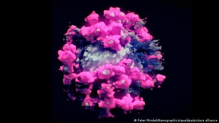
The real deal
And this is what the novel coronavirus actually looks like. Each SARS-CoV-2 particle is about 80 nanometers in diameter. Each particle contains a ball of RNA, the virus’s genetic code. That is protected by spike protein, the protusions that gave the virus its name. SARS-CoV-2 is a member of the coronavirus family, which includes the viruses responsible for SARS and MERS. More on that later.
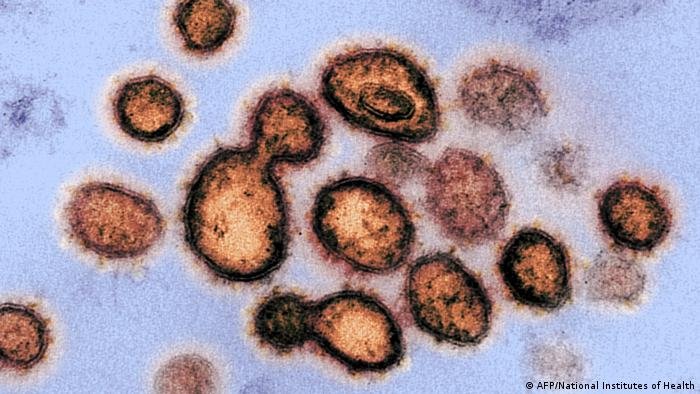
An airborne virus
SARS-CoV-2 particles are transferred through droplets and aerosols that a person emits when they breathe, cough or talk. That’s why face masks have become ubiquitous during the pandemic: Health authorities recommend citizens wear them to stop the spread of the virus. It can also be transmitted via contaminated surfaces.
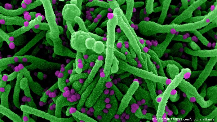
Entering human cells
SARS-CoV-2 uses its spike proteins to bind with a protein on the surface of cells. That sets off chemical changes, which allow the virus’s RNA to enter the cell (green in this image). The virus then forces the cell to make copies of its RNA. A single cell can produce tens of thousands of new virus particles (purple in this image) like this, which then infect other healthy cells.
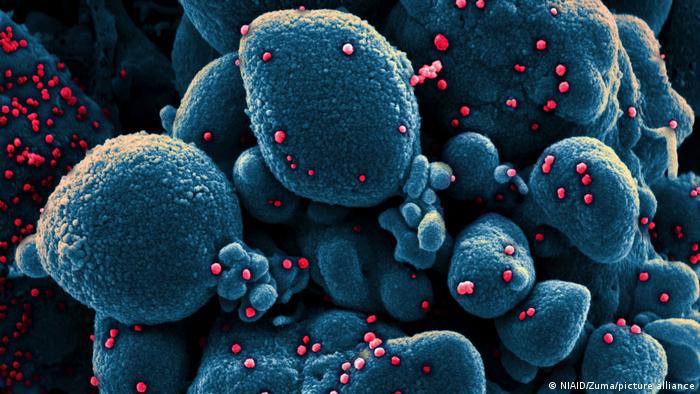
New to humans
Another electron microscope image of a cell (blue) heavily infected with SARS-CoV-2 particles (red). The virus behind our current pandemic isn’t too different from viruses like the ones causing the flu or common cold. But before 2019, human immune systems hadn’t seen this particular strain before, which is why no one had built up immunity.
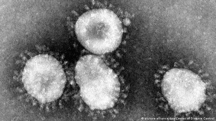
SARS-CoV: The first coronavirus outbreak of the 21st century
The first time this century that humanity came in contact with a coronavirus was in China in 2002. In March 2003, the WHO issued a global alert warning of atypical pneumonia spreading quickly. SARS, or severe accute respiratory syndrome, spread to roughly 30 countries, but not all of these recorded deaths. The WHO declared the epidemic contained in July 2003.
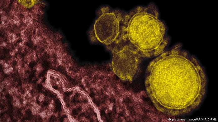
MERS-CoV, another coronavirus family member
In 2012, researchers discovered MERS-CoV, a novel coronavirus, after genome sequencing of samples from people who had fallen ill with a new flu-like illness. This illness came to be known as MERS, or Middle East respiratory syndrome, after where the first outbreak occurred. It is less infectious than COVID-19. Transmission usually occurs among family members or in healthcare settings.
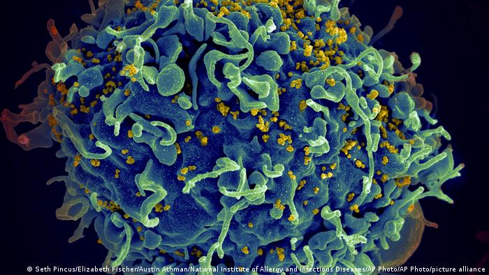
HIV: The other pandemic
The HI-virus (here in yellow), attacks the immune system, for examples T-cells (here in blue). Like SARS-CoV-2, it’s an RNA-based virus. If left untreated, it’ll weaken the immune system until it can’t fight infections anymore. HIV is transmitted through bodily fluids like semen or blood. There’s no vaccine, but there’s medication that brings down the viral load and stops AIDS from breaking out.
https://www.dw.com/en/covid-sars-cov-2-and-other-viruses-in-pictures/g-59901123